|
What We Use:
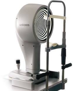
ANTARES CORNEAL TOPOGRAPHER The Antares instrument is a fully featured multi-functional corneal topographer. Antares has dedicated software designed to help in the detection and analysis of Dry Eye. The topography function provides information about the curvature, elevation and refractive power of the cornea. It also provides many parameters to aid in the diagnosis and monitoring of the corneal surface. |
|
Advanced Tear-film Analysis
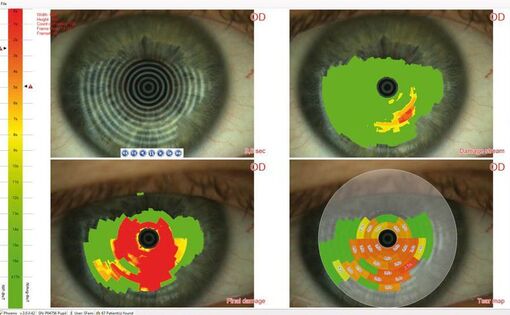
One common cause of dry eye symptoms is that the film of tears over the eye can break down or evaporate more quickly than it should, causing dry spots of unprotected cornea, conjunctiva, & other structures. This advanced, tear-film analysis is an expedient, clinical way to evaluate the severity of dry eye & help determine if treatment is working for you by measuring how long the tear-film on the eye stays unbroken. The photo to the right shows an example of the test results with a color-coded map of the tear film on the eye. Meibographer
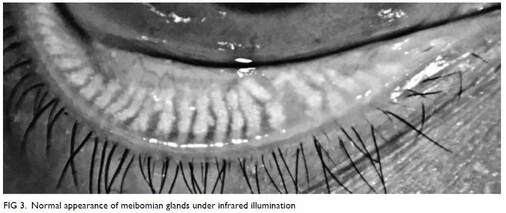
A meibographer uses infrared light to better highlight the meibomian glands within the eyelid. These glands secrete the lipid portion of the natural tear film that protects the eye and dysfunctions with these glands are considered a leading cause of dry eye. These glands can become clogged or can atrophy with age, contact lenses overuse, environmental factors, hormonal imbalance, and more. This scan allows the doctor to diagnose issues and monitor the progress of treatment. Fluorescein Imaging
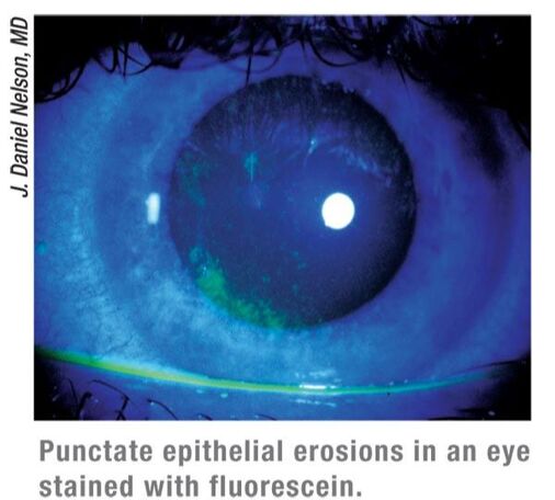
Dry eye syndrome can have negative effects on the outer surface of the eye. In this test, an orange dye called fluorescein is placed with a dropper onto the ocular surface & fluorescein uptake is measured & graded under cobalt blue light. This helps the doctor to detect if any ocular surface changes associated with insufficient tear film & excess dryness has occurred by helping visualize devitalized cells and tissues. Dry Eye Report
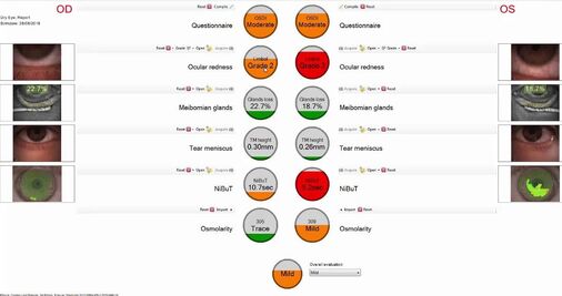
Based on the patient-reported Ocular Surface Disease Index questionnaire (OSDI) and several other tests including the TBUT, the doctor can tell what level of dry eye is present. This allows her to more accurately treat the condition and monitor the treatment's progress. The image to the right shows an example of the final report with green, orange, and red to show levels from normal to severe on each test result. |
